
Day|Week

 Mums stage breastfeeding flash mob
 Moscow “spider-man” climbs Chinese skyscraper
 Top 10 international destinations for Chinese millionaires
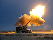
 New-type self-propelled gun fires in drill
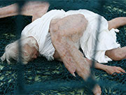
 Chinese artists create 'fall of an angel'
 A glimpse of China's Zijinshan gold & copper mine
 Hot figure show in SW China
 Evolution of Chinese beauties in a century