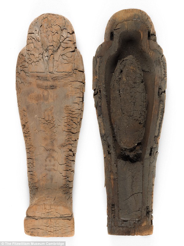
The landmark discovery was made by experts at the Fitzwilliam Museum in Cambridge who examined a wooden coffin donated to it by archaeologists in 1907.
Tutankhamun's tomb contained two small foetuses that had been mummified and placed in individual coffins.
But these infants were both significantly more developed, at about 25 weeks and 37 weeks into gestation.
Julie Dawson, head of conservation at the Fitzwilliam Museum said: 'Using non-invasive modern technology to investigate this extraordinary archaeological find has provided us with striking evidence of how an unborn child might be viewed in ancient Egyptian society.
'The care taken in the preparation of this burial clearly demonstrates the value placed on life even in the first weeks of its inception.'
Curators at the Fitzwilliam made the discovery, during their research for their bicentennial exhibition 'Death on the Nile: Uncovering the Afterlife of ancient Egypt.'
The tiny coffin, just 44 cm long, was excavated at Giza in 1907 by the British School of Archaeology and came into the collection at the Fitzwilliam Museum the same year.
It is a perfect miniature example of a wooden coffin of the ancient Egyptian late period dating from 664-525 BC.
The landmark discovery was made by experts at the Fitzwilliam Museum in Cambridge who examined a wooden coffin donated to it by archaeologists in 1907. Pictured is the open coffin with its contents. Although the coffin has deteriorated, it is clear that the wood was carefully carved in painstaking detail
The lid and box are both made from cedar wood.
Although the coffin has deteriorated, it is clear that the wood was carefully carved on a painstakingly small scale and decorated.
This gave the curators at the Fitzwilliam the first clear indication of the importance given to the coffin's contents at this time in ancient Egyptian society.
The diminutive wrapped package inside was carefully bound in bandages, over which molten black resin had been poured before the coffin was closed.
For many years it was thought that the contents were the mummified remains of internal organs that were routinely removed during the embalming of bodies.
Examination using X-ray imaging at the Fitzwilliam Museum was inconclusive.
It was decided to micro CT scan the tiny bundle at Cambridge University's Department of Zoology.
The cross-sectional images this produced gave the first pictures of the remains of a tiny human body held within the wrappings, which remain undisturbed.
Dr Tom Turmezei, former consultant radiologist at Addenbrooke's Hospital in Cambridge collaborated with the Fitzwilliam Museum, alongside Dr Owen Arthurs, academic consultant paediatric radiologist at Great Ormond Street Hospital, London.
 |  |
 Beijing Style: ready for bare legs
 Century-old station sees railyway evolution
 Amazing scenery of Xisha Islands
 Enthusiasts perform Kung Fu at Wudang Mountain
 Stunning photos of China's fighter jets in drill
 Monk's mummified body to be made into a gold Buddha statue
 Asia's longest and highest suspension bridge to open to traffic
 China's first interactive robot looks like a beauty
 Vietnamese Su-30 fighters fly over Nanwei Island in South China Sea
 Top 20 hottest women in the world in 2014
 Top 10 hardest languages to learn
 10 Chinese female stars with most beautiful faces
 China’s Top 10 Unique Bridges, Highways and Roads
 Caution urged in overseas M&A deals
 Activist’s cyber hunt violates moral codes
 China's radical leftists call for vocal critics of the Party to be jailed for treason
 Step aside Twiggy – more and more Chinese people are embracing a healthy body image
Day|Week

