Tue,Aug 12,2014
 Amphibious armored vehicle unit conducts open sea drill
 Water relay in Henan
 Ethnic culture feasts eyes of travelers
 80 security dogs assembled in Nanjing police dog training base
 Graffiti artists paint on street walls in Xinjiang
 Story of ceramic artist Zhang Lingyun
 Magic summer night dream in Hongyuan
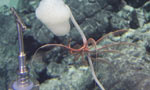
 Incredible creatures in headwaters drainage region of Lancang River
 The future of rock n' roll seen in young rockers in China
 Magnificent Yanziya Cliff
WASHINGTON, Aug. 11-- U.S. researchers said Monday they have created a three-dimensional brain-like tissue that functions like and has structural features similar to tissue in the rat brain and that can survive in the lab for more than two months.
The brain-like tissue, described in the U.S. journal Proceedings of the National Academy of Sciences, may offer new options for studying brain function, disease and trauma, and treatment.
The key to generating the tissue was the creation of a novel composite structure that consists of two biomaterials with different physical properties: a spongy scaffold made out of silk protein and a softer, collagen-based gel, researchers at the Tufts University said.
They first cut the spongy scaffold into a donut shape and populated it with rat neurons. Then, the researchers filled the middle of the donut with the collagen-based gel, which subsequently permeated the scaffold to encourage neuron growth.
In just a few days, the neurons clustered within the pores of the scaffold, forming long-lasting networks in the gels that resembled the complex circuitry of the brain in the rat.
"With the system we have, you can essentially track the tissue response to traumatic brain injury in real time," said Professor David Kaplan of the Tufts University who led the research efforts to develop the tissue.
"Most importantly, you can also start to track repair and what happens over longer periods of time."
The researchers were able to use the tissue model to examine multiple post-injury effects, including cellular damage, electrophysiological activity and neurochemical changes.
For example, when a weight was dropped on the model tissue to simulate a traumatic brain injury, the tissue released high levels of the chemical glutamate, a neurotransmitter known to be emitted by cells following brain damage, the researchers said.
The tissue also showed transient electrical hyperactivity consistent with post-trauma responses observed in vivo, they said.
Kaplan emphasized the importance of the brain-like tissue's longevity for studying other brain disorders.
"The fact that we can maintain this tissue for months in the lab means we can start to look at neurological diseases in ways that you can't otherwise because you need long timeframes to study some of the key brain diseases," he said.
Previously, scientists have attempted to grow neurons in 3D gel environments, yet these gel-based tissue models don't live long and fail to yield robust, tissue-level function.
 Beijing policewomen posters become a hit
 Armored regiment trains on the sea
 Children spend 'Father's Day' with dads at work
 'Pan Da' appear in Shanghai World Financial Center
 Champions take selfies on podium
 National Fitness Day celebrated around China
 Traditional culture colors summer vacation
 Young athletes fighting for their dreams
 68 meters high thermometer in Shanxi, called ‘fighter’ of thermometers
 22-year-old veteran travels around China
 Night scenery of pagoda forests
 China suffers from hot summer
 48 hours after super Typhoon Rammasun
 German pianist plays mid-air ‘magic carpet’ show over Munich Airport
 China's manned deep-sea submersible conducts dive in Pacific Ocean
Day|Week|Month